
Figure 3.
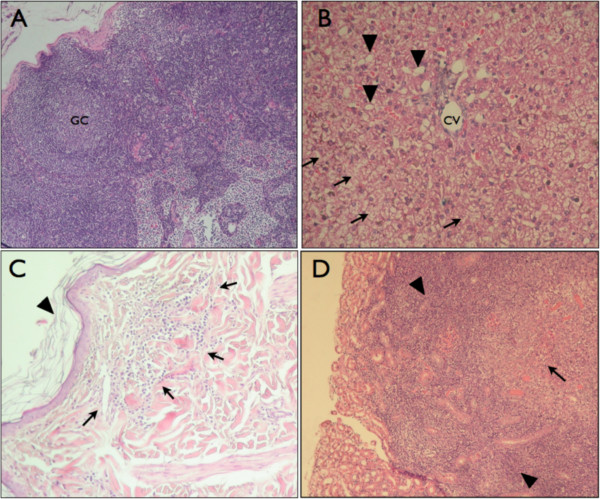
Photomicrographs of hematoxylin eosin stained sections of tissues from infected dogs. (A) Cortical zone of the prescapular lymph node. GC: Germinal Centre. Magnification 10×. (B) Liver section, magnified 20×. Hepatocytes present hydropic degeneration (arrowhead). Necrotic cells are scattered throughout parenchyma (arrows). CV: Central vein. (C) Epidermis section magnified 20×. Inflammatory infiltrate is present in the dermis (arrows) and epidermis integrity is lost by orthokeratotic hyperkeratosis (arrowheads). (D) Kidney section magnified 10×. Severe inflammatory infiltrate is observed in the interstitium (arrowheads). Severe lymphocytic and histiocytic inflammatory infiltrate is around necrotic zone (arrow).