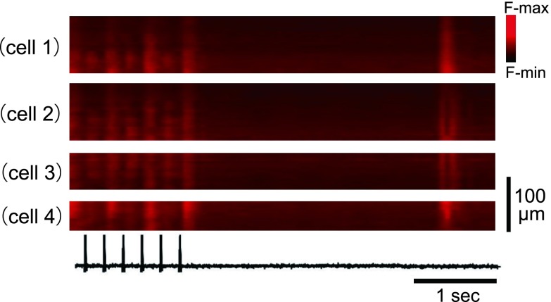
Fig. 6. .
Absence of the spontaneous Ca2+ waves after burst pacing of the left atrium showing beat-to-beat Ca2+ alternans. X-t images of four different cells. Electrical stimuli are shown on the bottom. Six non-uniform Ca2+ transients were evoked by burst pacing at 5 Hz. About 300 milliseconds after pacing, one spontaneous Ca2+ transient occurred showing relatively uniform wavefronts for individual cells.

