
Figure 1.
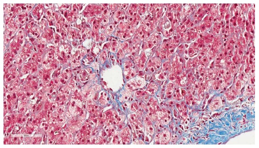
Histopathology of fibrosing cholestatic hepatitis C. Histopathology of fibrosing cholestatic hepatitis C demonstrating periportal sinusoidal and pericellular “chicken wire” fibrosis (trichrome, image magnification × 40) (Courtesy of Carl Jacobs, MD, Department of Pathology, Carolinas Medical Center, Charlotte, NC, United States).