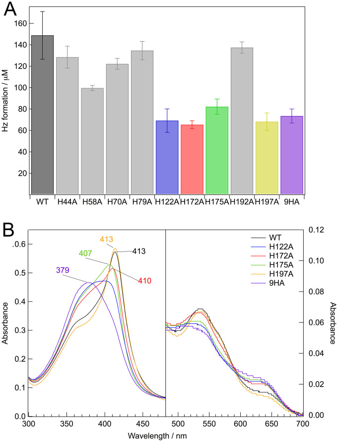
Figure 3. Hz formation activity and electronic absorption spectra for HDP and its mutant proteins.
(A) Hz formation assay for wild-type HDP (WT) and mutant proteins. The assays of Hz formation were carried out for 15 min at 37°C as described in the Methods section. Data are expressed as the mean and standard deviation of at least four independent experiments. (B) Electronic absorption spectra of heme-bound wild-type HDP (WT) and mutant proteins. The sample concentration was approximately 40 μM in 50 mM MOPS–NaOH, pH 7.0, at room temperature. The path-length of the cell was 0.2 cm.

