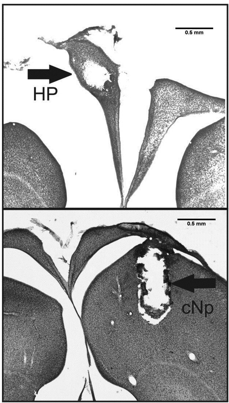
Figure 1.
Representative coronal sections from an individual with a microdialysis probe in the left HP (top panel) and an individual with a microdialysis probe in the cNp (bottom panel). Arrows point to the damage caused by the microdialysis probe (Note: the HP becomes separated from the underlying nidopallium at the lateral ventricle when tissue is slide mounted).

