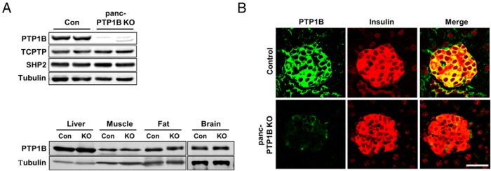
Figure 1.

PTP1B deletion in the pancreas. A, Upper panel, Immunoblots of PTP1B expression in primary islets from PTP1Bfl/fl (control [Con]) and panc-PTP1B KO female mice fed an HFD at 28 weeks of age. Lysates were also probed for T-cell PTP (TCPTP), Src homology phosphatase 2 (SHP2), and tubulin. Lower panel, Immunoblots of PTP1B expression in liver, muscle, fat, and brain of control and panc-PTP1B KO mice. Lysates were also probed for tubulin. B, Confocal images of pancreas sections from female control and panc-PTP1B KO mice fed regular chow at 20 weeks of age stained for PTP1B (green) and insulin (red) and merged. Scale bar, 100 μm.
