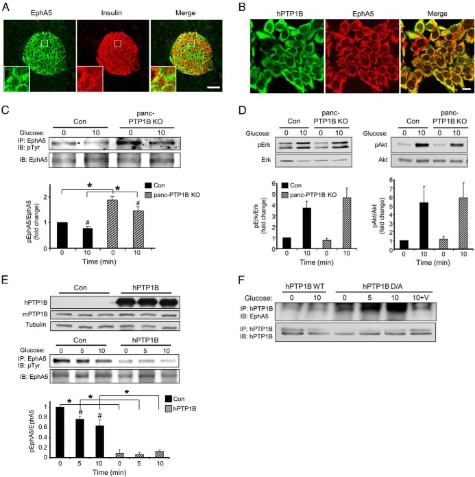
Figure 4.
PTP1B regulates EphA5 tyrosyl phosphorylation in islets and MIN6 β-cells. A, Confocal images of pancreas sections stained for EphA5 (green) and insulin (red) in control female mice fed regular chow for 3 months. Boxed areas are magnified in the insets. Scale bar, 100 μm. B, Confocal images of MIN6 β-cells stained for hPTP1B (green) and endogenous EphA5 (red). C, Primary islets from control (Con) and panc-PTP1B KO mice fed an HFD were starved then stimulated with 25mM glucose for 10 minutes. EphA5 immunoprecipitates were immunoblotted for phosphotyrosine (pTyr) and EphA5. Bar graph represents normalized data from 3 mice per group. Data are presented as mean ± SEM. *, Significant differences between groups; #, significant differences between basal and glucose-stimulated conditions within a group. D, Islet lysates were immunoblotted for pERK, ERK, Akt, and Akt. Data are presented as mean ± SEM; n = 4. E, EphA5 tyrosyl phosphorylation in MIN6 β-cells overexpressing hPTP1B. Cell lysates were immunoblotted for hPTP1B, mPTP1B, and tubulin (top panel). EphA5 was immunoprecipitated from control and MIN6 β-cells overexpressing hPTP1B under basal (0) and glucose-stimulated (5 and 10 minutes) conditions and then immunoblotted for pTyr and EphA5. Data of pEphA5/EphA5 are presented relative to control (0) and as mean ± SEM; n = 3. *, Significant differences between groups; #, significant differences between basal and glucose-stimulated conditions within a group. F, MIN6 β-cells were transfected with WT hPTP1B and substrate-trapping mutant D/A. Cells were starved and then stimulated with glucose (25mM) and lysed as indicated in Materials and Methods with and without vanadate treatment. hPTP1B was immunoprecipitated and then immunoblotted for endogenous EphA5 and hPTP1B. Abbreviations: IB, immunoblot; IP, immunoprecipitation.

