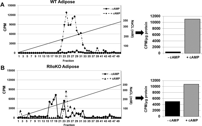
Figure 3.
DEAE-cellulose column separation and PKA activity assay of inguinal adipose from HFD-fed WT (A) and RIIαKO (B) mice showed substantially increased basal kinase activity in the mutant mice. Line graphs show kinase activity in HPLC-separated fractions with and without added cAMP (5 μM). Type I elutes between 40 and 80 mM (fractions 20–25), type II elutes between 120 and 200 mM (fractions 31–35), and free C subunit elutes just before the type I holoenzyme. Bar graphs show kinase activity (per microgram of protein) in lysate prior to HPLC separation (cumulative kinase activity with and without added cAMP).

