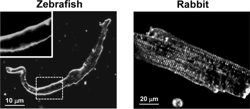
Figure 1. Morphology of zebrafish and rabbit ventricular myocytes.

Confocal 2-D images of ventricular myocytes from zebrafish (left panel) and rabbit (right panel) labeled with the voltage-sensitive fluorescent dye Di-8-ANEPPS. The left-top panel is an enlarged area marked with the dashed white line, to highlight the absence of the t-tubular network in the zebrafish ventricular myocytes.
