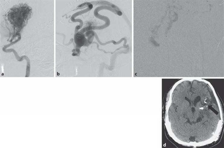
Fig. 4.
A 39-year-old male presented with SAH related to a large AVM involving the left frontal lobe, left basal ganglia and left temporal lobe. a Lateral view, left ICA injection – arterial phase. b Left ICA injection – venous phase. c Subtracted microcatheter injection of feeding pedicle. d NBCA in nidus (arrow) on unenhanced CT image.

