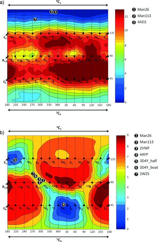
Figure 3.

Conformational free-energy landscapes (FELs, Mercator projection) of isolated isofagomine (1; a) and protonated mannoimidazole (2; b), contoured at 1 kcal mol−1. FELs have been annotated with the conformations of isofagomine-type (for a) and mannoimidazole-type (for b) inhibitors which have been observed on-enzyme. a) 1: 3 bound to GH26 CjMan26C (this work); 2: 3 bound to GH113 AaManA (this work); 3: α-Glc-1,3-isofagomine bound to BxGH99 (PDB code 4AD2).17 b) 1: 4 bound to GH26 CjMan26C (this work); 2: 4 bound to GH113 AaManA (this work); 3: 1 bound to GH2 BtMan2A (PDB code 2VMF);18 4: 1 bound to GH47 CkMan47 (PDB code 4AYP);11 5: 1 bound to GH38 DmGManII (PDB code 3D4Y) in half-chair conformer;19 6: 1 bound to GH38 DmGManII (PDB code 3D4Y) in boat conformer (this work); 7: 1 bound to GH92 BtMan3990 (PDB code 2WZS).20
