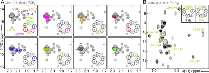
Figure 3.

Methyl-group labeling. A) Methionine methyl-group spectra of the LSm1–7 complex. Top left: the methyl TROSY spectrum of an LSm1–7 complex in which all the LSm proteins are NMR active. This spectrum can be deconvoluted into seven simplified spectra that contain only a singly NMR-active LSm protein (other panels). B) Methyl TROSY spectra of Ile-δ1 labeled LSm2–8 complexes. The spectrum of the fully isoleucine labeled spectrum (gray) is simplified by labeling only the LSm5, LSm6, and LSm7 proteins (black) or only the LSm5 protein (olive).
