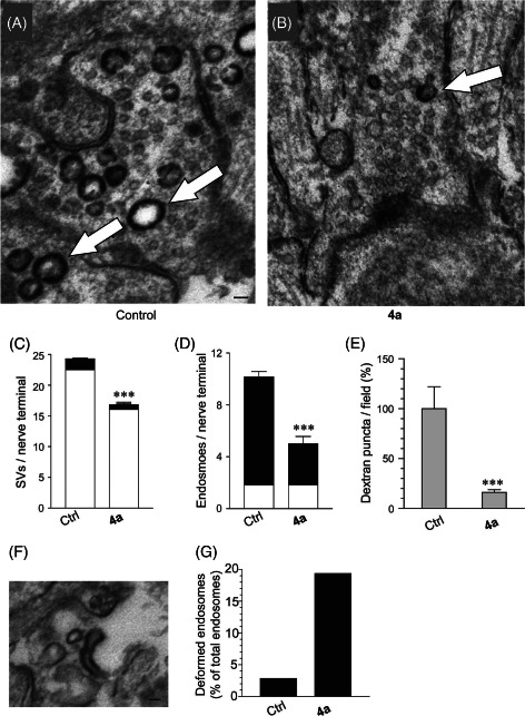
Figure 4.

Dyngo compound 4a inhibits both CME and ADBE. Uptake of HRP in response to electrical stimulation (80 Hz for 10 seconds) was examined in CGNs. Neurons were preincubated with or without 30 μM 4a for 15 min before and during stimulation. Panels show typical electron micrographs of CGNs after HRP labeling and fixation, either in the absence (A) or presence of 4a (B). HRP‐labeled endosomes are indicated by arrows. Scale bar represents 100 nm. Panel (C) shows quantitation of the number of synaptic vesicles observed in control and 4a‐treated samples. Synaptic vesicles were either unlabeled (white bar) or HRP labeled (black bar). 4a treatment significantly reduced the number of unlabeled and labeled synaptic vesicles, demonstrating an inhibition of CME. Panel (D) shows quantification of labeled and unlabeled endosomes. 4a inhibited the number of HRP‐labeled endosomes, demonstrating an inhibition of ADBE. E) 4a (30 μM) inhibited uptake of fluorescent dextran in CGNs electrically stimulated at 80 Hz, further suggesting inhibition of ADBE. F and G) In electron micrographs, 4a treatment also increased the appearance of deformed HRP‐labeled endosomes, such as that shown in (F). Scale bar represents 50 nm. Quantification of the number of deformed endosomes for each condition is shown in (G), presented as % of total endosome number. All data are means ± SEM, ***p < 0.001, Student's t‐test.
