Figure 1.

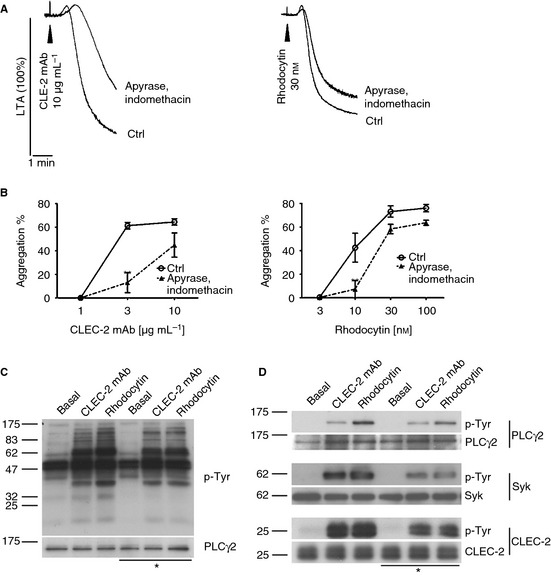
CLEC‐2‐dependent activation of mouse platelets is partially mediated by ADP and TxA2‐. Washed platelets (2 × 10⁸ per mL) were stimulated with 10 μg mL−1 CLEC‐2 mAb or 30 nm rhodocytin in the presence or absence of 2 U mL−1 apyrase and 10 μm indomethacin. Representative light transmission aggregometry (LTA) traces are shown (A). Aggregation after 5 min was plotted as mean ± SEM (n = 4–6). Statistical differences were evaluated by two‐way anova and Bonferroni post‐test (**P < 0.01) (B). Washed platelets (4 × 10⁸ per mL) incubated with 10 μm lotrafiban were stimulated with 10 μg mL−1 CLEC‐2 mAb and 30 nm rhodocytin for 3 min in the presence (*) and absence of 2 U mL−1 apyrase and 10 μm indomethacin. Whole cell lysate was analyzed by SDS‐PAGE (C) and immunoprecipitation of PLCγ2, Syk and CLEC‐2 (D). Blots were probed with anti phospho‐tyrosine (p‐Tyr) antibody and reprobed for equal loading control (n ≥ 3) (C&D).
