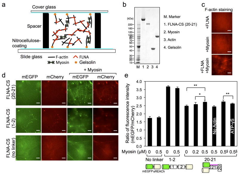
Figure 4. Myosin-dependent conformational changes of purified FLNA-CS in actin networks.
(a) Actin networks crosslinked with FLNA-CS are internally stressed by myosin motors. Actin filaments are anchored to glass surface through gelsolin coated on the surface. (b) Coomassie blue stain of SDS-PAGE gel of purified proteins used in this study. (c) 10 μM F-Actin, crosslinked with 0.1 μM FLNA or not, was internally stressed by 0.5 μM myosin. F-actin is stained with Alexa Fluor® 568 phalloidin. (d) Fluorescence images of mCherry-FLNA-CS (0.1 μM) embedded in actin filaments (10 μM) in the presence or absence of myosin (0.5 μM). Ratio of fluorescence intensities of mCherry and mEGFP was calculated and plotted in panel e. Scale bar = 10μm. (e) Error bars represent SD, n ≥ 4 independent experiments. §These two experiments were independently performed and compared. Statistical significance was determined by a two-tailed t test (*P<0.05, **P<0.005).

