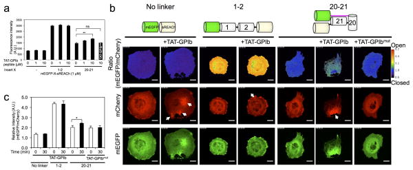
Figure 5. A cell-permeable high affinity ligand (TAT-GPIbα) for repeat 21 of FLNA induces conformational changes in the FLNA PQ-FRET probe (FLNA-CS) expressed in COS-7 cells.
(a) Effect of increasing amounts of TAT-GPIbα peptide (0 – 10 μM) or TAT-GPIbα mutant peptide (10 μM) on the fluorescence intensity of purified mEGFP-sREACh constructs containing no linker, linked together with FLNA repeats 1–2, or linked together with repeats 20–21 (1.0 μM each). Fluorescence was measured in a microplate reader with excitation/emission wavelengths of 458/511 nm (mEGFP). (b) Fluorescence images of mCherry and mEGFP of FLNA-CS expressed in COS-7 cells before and after treatment with TAT-peptides. Ratio images and Ratio color scale (mEGFP/mCherry) were generated using Ratio Plus plugin (ImageJ). The cells were treated with 100 μM cell-permeable TAT-GPIbα peptide or TAT-GPIbα mutant peptide. Arrows highlight foci induced by the cell-permeable TAT-GPIbα peptide. Note that the ratio values at the foci are not quantitative because the fluorescence intensity was saturated. Scale bar = 20μm. The corresponding movies are shown in Supplementary movie 1. (c) The ratio of fluorescence intensities of mEGFP versus mCherry are plotted before and after treatment with the cell-permeable peptides. Error bars represent SD (n=3). *P<0.05, **P<0.005; ns, not significant by a two-tailed t test.

