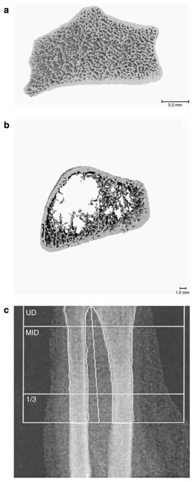
Figure 3. Example images from techniques to evaluate bone structure.

Representative HR-pQCT images from a healthy patient (a); a patient with ESKD (b); and a DXA image from the same patient with ESRD (c). HR-pQCT of the radius of the patient with ESKD demonstrates cortical thinning and extreme trabecular dropout. In comparison with DXA imaging of the same bone, HR-pQCT provides superior resolution with visualization of both trabecular and cortical bone compartments.
