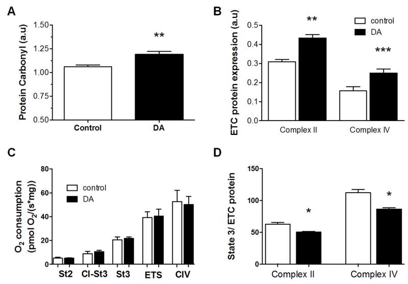
Figure 4.
Comparison of oxidative damage and mitochondrial content and function in brains of zebrafish exposed for 18 weeks to domoic acid and time-matched vehicle controls. (A) Protein carbonyl levels were significantly higher in exposed fish compared to controls. (B) Expression of complex II (30 kD subunit) and complex IV (subunit IV) were both significantly higher in exposed fish compared to controls. (C) Oxygen flux per unit brain tissue at five different measures of mitochondrial respiration (St4 = state 4, C1-St3 = Complex I State 3, St3 = Complex I & II State 3, ETS = Fully uncoupled respiration, and CIV = Complex IV) was not significantly different between control and exposed fish. (D) Maximal state 3 respiration (ADP, plus succinate and glutamate/malate) was normalized to complex ll and complex IV protein content. Flux per content was reduced in exposed fish compared to controls. n=6–11 for control and exposed treatments, mean ± SEM. *-P<0.05, **-P<0.01, ***-P<0.0001 relative to controls.

