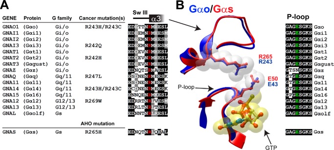
FIGURE 1.
Identification of disease-linked mutations in a conserved arginine of Gα proteins. A, left, list of cancer somatic mutations in the position corresponding to Gαo Arg-243 in different Gα subunits. Mutations were identified by bioinformatics searches as described in “Experimental Procedures.” The Gαs R265H mutation found in AHO is shown below. Right, alignment of sequences flanking the conserved arginine (in red) from all 16 human Gα subunits. B, left, overlay of the P-loop and SwIII/α3 of active Gαo (blue, PDB: 3C7K) and active Gαs (red, PDB: 1AZT) with a ball and stick representation of Arg-243/265 and Glu-43/50. Nucleotide is shown in yellow. Right, alignment of the P-loop sequence from all 16 human Gα subunits.

