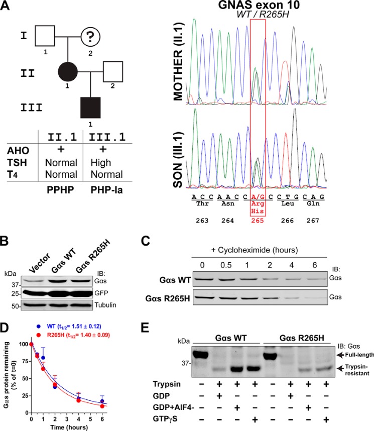
FIGURE 2.
Gαs R265H is a bona fide loss-of-function mutation in AHO. A, left, pedigree of the family affected by the Gαs R265H mutation. Squares, male; circles, female; black symbols, AHO. The question mark indicates a deceased patient who had external signs of AHO but was not formally diagnosed before death. The table below summarizes clinical features of the two patients with confirmed AHO. Right, sequence analysis of a fragment of Gαs exon 10 displaying a heterozygous substitution of A for G in codon 265. B, Gαs R265H protein expression levels in mammalian cells are similar to Gαs WT. HEK293 cells were transfected with equal amounts of plasmids encoding for Gαs WT or Gαs R265H, lysed, electrophoresed, and immunoblotted with the indicated antibodies. Equal Gαs WT and Gαs R265H plasmid transfection is verified by the equal levels of GFP expression, which is driven by the same promoter as Gαs via an IRES. One experiment representative of three is shown. C and D, Gαs R265H protein half-life in mammalian cells is similar to Gαs WT. HEK293 transfected with Gαs WT or Gαs R265H were treated with cycloheximide (20 μg/nl) for the indicated times, lysed, and immunoblotted with Gαs antibodies. A representative blot is shown in C, and the quantification of two independent experiments (mean ± S.D.) fitted to a single exponential decay curve is shown in D. E, Gαs R265H expressed in mammalian cells has impaired nucleotide binding as assessed by trypsin protection assays. HEK293 cells expressing Gαs WT or Gαs R265H were lysed and incubated in the presence of GDP, GTPγS, or GDP plus AlCl3/NaF (AlF4−) before treatment with trypsin as described in “Experimental Procedures.” Samples were subjected to SDS-PAGE and immunoblotted with the indicated antibodies. One experiment representative of three is shown.

