FIGURE 2.
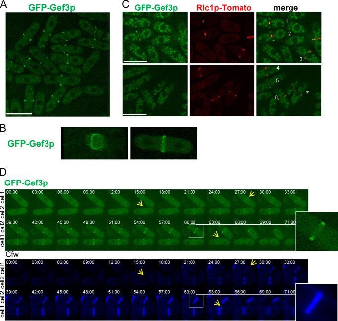
Gef3p localizes as a medial ring. A, cells producing GFP-Gef3p (SM217) were synchronized with 12.5 mm hydroxyurea (HU) for 2.5 h, washed 3x, and photographed 1 h after release into fresh medium. Bar, 10 μm. B, cells expressing GFP-Gef3p were imaged for GFP fluorescence using confocal three-dimensional microscopy. Frontal and lateral views of the GFP-Gef3p septum are shown (see supplemental Movies 1 and 2). C, cells (SM267) expressing GFP-Gef3p and Rlc1p-tdTomato were imaged for GFP and RFP fluorescence. During ring contraction, most Gef3p remains as an external ring (cells 1 and 2). Cells 3, 4, and 7 exhibit myosin but not Gef3p staining, whereas cell 5 shows just the opposite. Bar, 10 μm. D, GFP-gef3+ cells were imaged by time-lapse microscopy. The elapse time is shown in minutes. The inset shows that the GFP-Gef3p ring splits into a double ring.

