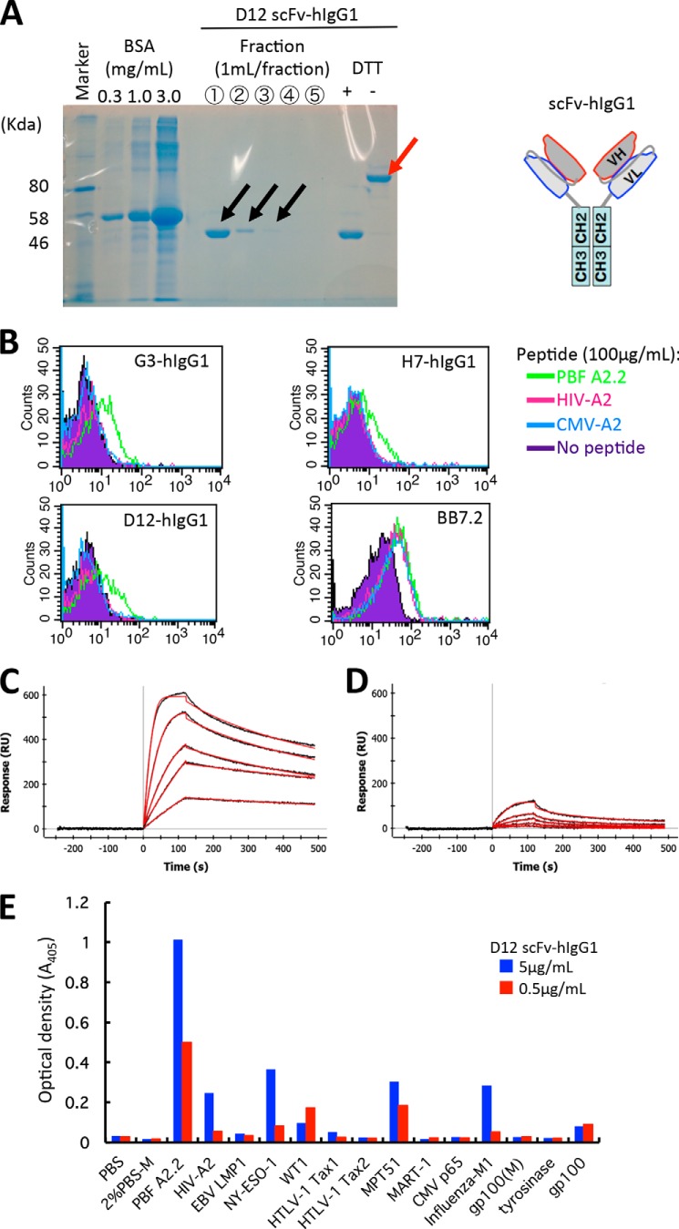
FIGURE 4.
The scFv clones could react with peptide PBF A2.2 presented by HLA-A*02:01 on antigen-presenting cells with strong affinity. A, soluble fractions of D12 scFv-hIgG1 after purification with Protein G are visualized by SDS-PAGE. The reduced monomer (black arrows) and oxidized dimer of scFv-hIgG (red arrow) of fraction 1 are indicated. B, FACS analysis of scFv-hIgG1. T2 cells were pulsed with the indicated peptides and stained with each scFv-hIgG1 at a concentration of 10 μg/ml. BB7.2 was used to detect expression of HLA-A2 molecules. C and D, surface plasmon resonance analysis. Biotinylated HLA-A*02:01·PBF A2.2 peptide complex (C) or HLA-A*02:01·HIV-A2 peptide complex (D) was immobilized on the sensor tip as the target. Serially diluted D12 scFv-hIgG1 was used as the analyte as described under “Experimental Procedures.” E, ELISA screening of the reactivity of D12 scFv-hIgG against various biotinylated HLA-A*02:01·peptide complexes. RU, response units; EBV, Epstein-Barr virus; HTLV-1, human T-lymphotropic virus type I.

