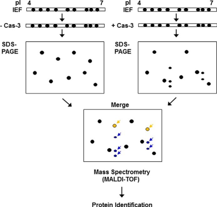
FIGURE 1.

A schematic view of the novel caspase-3 substrate screening method. After the IEF step, the strips were incubated in a caspase-3 activation buffer with or without an empirically predetermined amount of the recombinant human caspase-3 (Cas-3). Subsequently, SDS-PAGE was performed, and the resulting gels were stained with Coomassie Brilliant Blue G-250. The separated protein spots were analyzed by using ProteomWeaver software system. Assuming that the cleaved forms of the putative caspase-3 substrates (indicated by blue arrows) appear below the uncleaved full-length forms (indicated by yellow arrows) on the merged gel, all of the spots indicated by arrows were subjected to MALDI-TOF mass spectrometry for identification.
