
FIGURE 2.
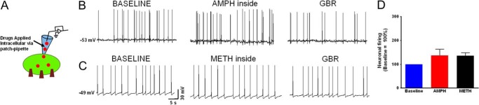
Intracellular amphetamine or methamphetamine less effectively modulates the firing rate of dopaminergic neurons. A, schematic depicts the experimental configuration showing DAT substrates were applied intracellularly. B and C, current clamp recording of midbrain dopaminergic neurons showing the spontaneous firing activity at the endogenous resting membrane potential when DAT substrates, amphetamine (1 μm, n = 4), or methamphetamine (1 μm, n = 5) were dialyzed into the neuron to bypass the forward transport step. At the resting membrane potential the spontaneous firing activity of the neurons immediately after achieving whole-cell configuration reflects the base-line firing activity of the neuron. GBR, GBR12935. D, intracellular amphetamine or methamphetamine equally but only modestly affected the firing rate of dopaminergic neurons (black and red bar). The bar graphs show the spontaneous firing activity of the neurons when amphetamine or methamphetamine was dialyzed into the neuron. The data are normalized to the base-line spontaneous firing rate of each experiment. The change in firing rate was blocked by extracellular application of a DAT antagonist, GBR12935 (1 μm).