FIGURE 6.
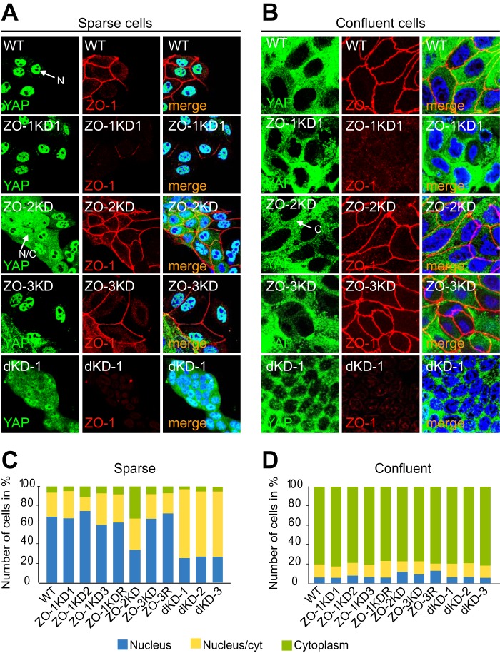
ZO-2 but not ZO-1 or ZO-3 is required for the nuclear localization of YAP in MDCK cells. A and B, immunofluorescent localization of Yap (green) and ZO-1 (red) in sparse (A) and confluent (B) MDCK WT cells, and cells depleted of ZO-1 (ZO-1KD1), ZO-2 (ZO-2KD), or ZO-3 (ZO-3KD). Merge images (merge) show nuclei labeled in blue by DAPI. C and D, composite histograms showing the percentage of cells with exclusively nuclear (blue), exclusively cytoplasmic (green), or mixed nuclear-cytoplasmic (yellow) localization (see legend) in the different clonal and rescue lines, cultured at either sparse (C) or confluent (D) density. N, N/C, and C in immunofluorescent panels indicate an example of nuclear, nuclear/cytoplasmic, and cytoplasmic localization, respectively.

