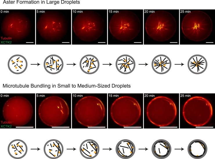
FIGURE 5.
Time course of confined motor-driven microtubule self-organization. Shown is motor-driven microtubule self-organization inside DOPC/DOPE/DOPG-monolayered droplets of different sizes. Also shown is a time series of characteristic images of motor-driven microtubule organization inside a large (85-μm diameter, top panel) and a medium-sized (30-μm diameter, bottom panel) lipid-monolayered droplet containing 40 μm Atto633-labeled tubulin (red) and 200 nm mCherry-kinesin-14 (green) in standard droplet buffer, as acquired with spinning disk confocal microscopy at 32 °C. Schematics of the observed microtubule (black) and motor (orange) organizations inside droplets (gray) are shown below the experimental data. Scale bars = 20 μm.

