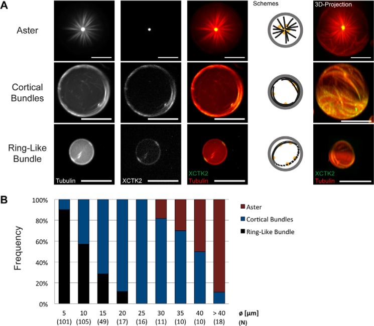
FIGURE 6.
Droplet size dependence of confined motor-driven microtubule self-organization. A, different categories of end states. Spinning disk confocal microscopy images of DOPC/DOPE/DOPG-monolayered droplets containing 40 μm Atto633-labeled tubulin (red in merge) and 200 nm mCherry-kinesin-14 (green in merge) in standard droplet buffer taken 25 min after reaction start are shown. Single channel and merged channel images of the equatorial droplet plane are shown as indicated. The schematics display the arrangements of microtubules (black) and the localization of the motor protein (orange) inside lipid-monolayered droplets (gray). The images on the right show three-dimensional projections. Scale bars = 20 μm. B, bar graph displaying the relative frequencies of the motor-organized microtubule organizations for different droplet diameters (Ø). Numbers in parentheses indicate the total count (N) of droplets analyzed for each size category.

