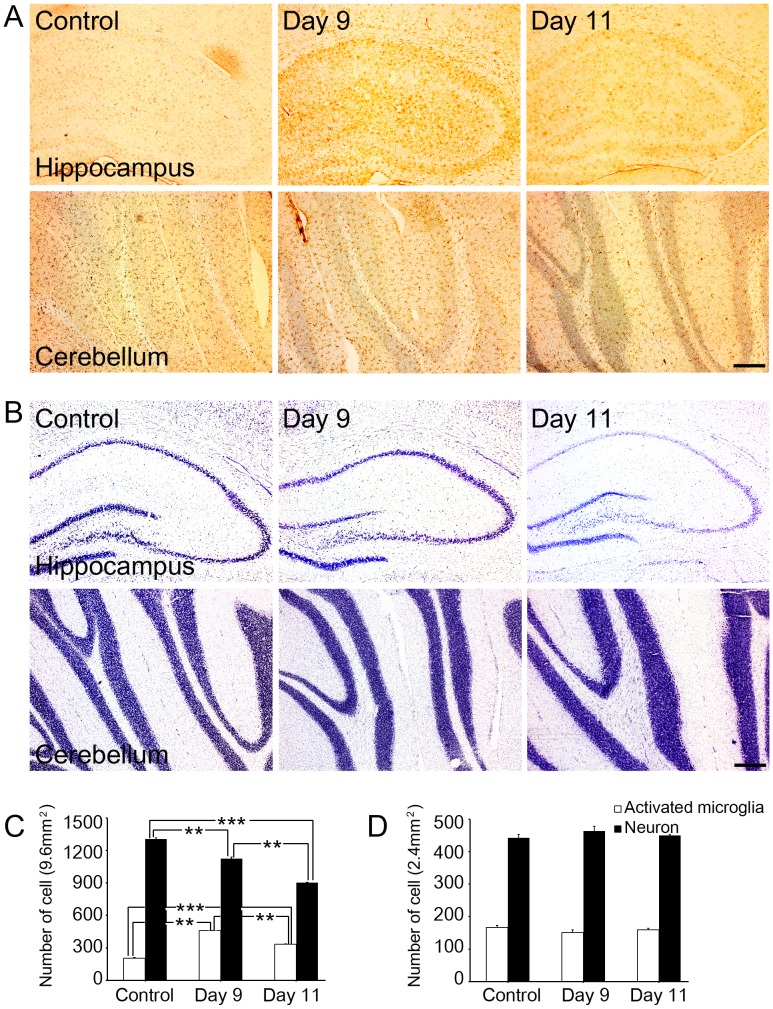
Figure 2. Distribution of activated microglial cells and neuron in the hippocampus of the control or rats exposed to binge-alcohol at days 9 and 11.
(A) OX-42-positive microglial cells were measured by immunohistochemical staining in control or alcohol-exposed rats brains. (B) Cresyl violet staining was used to detect decreasing neuronal cell number in the hippocampus of alcohol-exposed rats. (C) Number of OX-42 positive and cresyl violet stained cells in the hippocampus in control or alcohol-exposed rats. (D) Number of OX-42 positive and cresyl violet stained cells in the cerebellum at control or alcohol-exposed rats (white bar; OX-42 positive cell (activated microglia), black bar; cresyl violet positive cell (neuron)). Scale bar = 200 µm. **; p<0.001, ***; p<0.0001.

