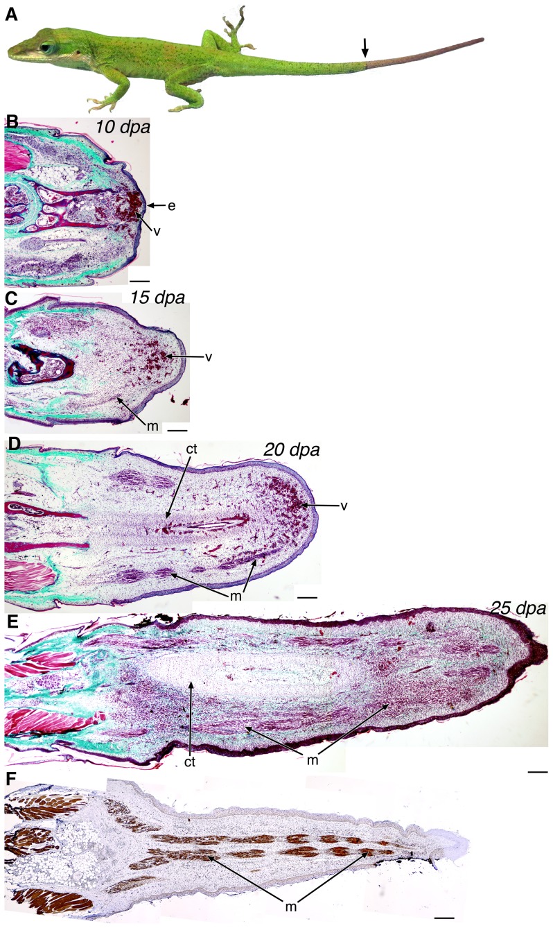
Figure 1. Overview of the stages of lizard tail regeneration.
(A) Anolis carolinensis lizard with a regenerating tail (distal to arrow). (B-E) Histology of the 10 dpa (B), 15 dpa (C), 20 dpa (D), and 25 dpa (E) regenerating tail by Gomori's trichrome stain, with which connective tissues and collagen stain green-blue, muscle, keratin, and cytoplasm stain red, and nuclei are black. (F) Immunohistochemistry of myosin heavy chain in a 25 dpa regenerating tail using the MY-32 antibody. e, wound epithelium; v, blood vessels; m, muscle; ct, cartilaginous tissue. Composites: B-F. Scale bars in black: 200 µm.

