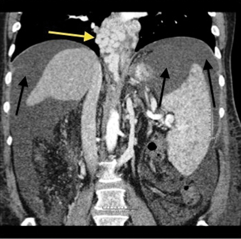
Figure 1.

Coronal contrast-enhanced computed tomography of the abdomen demonstrates avidly enhancing, markedly tortuous, and dilated varices surrounding the lower esophagus (yellow arrow). There is also significant perihepatic and perisplenic ascites (black arrows). The spleen is enlarged secondary to portal hypertension.
