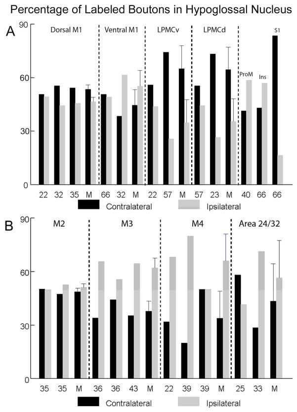
Figure 6.
Estimated percentage of labeled boutons in the ipsilateral and contralateral hypoglossal nucleus following injections of high-resolution anterograde dextran tract tracers in lateral (A, top) and medial (B, bottom) cortical regions. Each individual SDM monkey case is identified on the abscissa as well as the mean (M) for each major cortical region (separated by the vertical dashed lines) when more than one animal case was available for study. The error bars on the mean for each major cortical projection represent standard errors when more than one case was available for study.

