Figure 6.
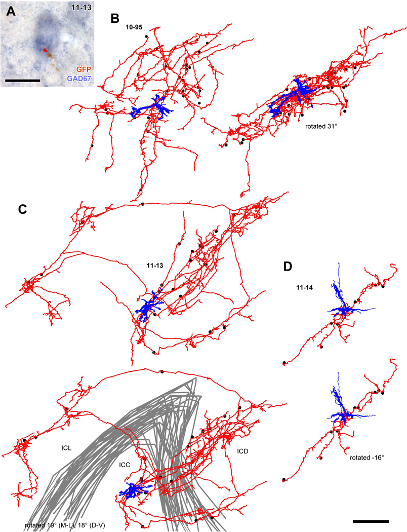
Spatial distribution of GAD67+ cell bodies receiving axosomatic contact from axons of a single glutamatergic neuron. (A) An example of GAD67+ neuron (blue) that received axosomatic contact (a red arrow) with a GFP+ axon (brown). Case 11-13. (B–D) Reconstructed dendrites (blue) and axons (red) of GFP+ neurons shown in Fig. 5. Dots indicate cell bodies of GAD67+ neurons that receive one or more axosomatic contacts. In case 10-95 (B), the axonal plexus became flatter when the cell was rotated 31° around the medio-lateral axis. In case 11-14 (D) the cell had a poorly arborized axon, and the GAD67+ cells aligned well after a rotation of −16° around the medio-lateral axis. In the case 11-13 (C), axon collaterals made two planar plexuses in the ICC and ICD, and one plexus in the ICL. The GAD67+ cells receiving the axosomatic contacts were located in the dorsal and lateral cortices as well as in the ICC. Gray lines indicate the border of the ICC. Scale bars: 20 µm (A) and 250 µm (B–D).

