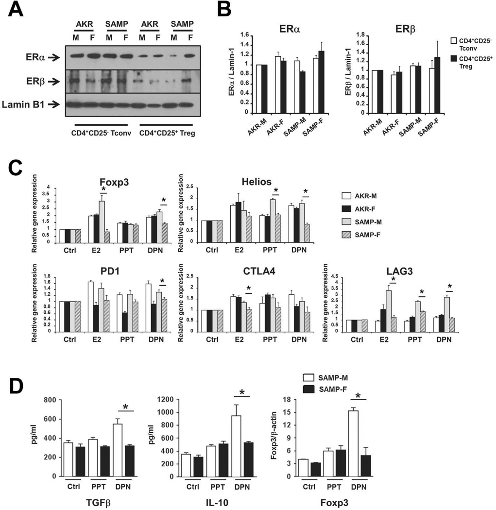
Figure 4. Estrogen signaling differentially affects Treg induction in SAMP-M vs. SAMP-F mice.
(A, B) CD4+CD25− Tconv and CD4+CD25+ Treg cells were isolated from spleens of indicated mice (10 weeks of age). Nuclear protein lysates were probed for ERα, ERβ, and Lamin-1 by Western blot. Representative images (A) and semi-quantitative densitometric analysis (B) are shown (n=3 total experiments, 3–5 pooled mice per group). (C) CD4+CD25− T cells were isolated from untreated, 10 week old mice and cultured in vitro with vehicle (Ctrl), E2, PPT, or DPN (10nM). mRNA specific for indicated genes was normalized to GAPDH and mean fold changes are expressed ± SEM relative to vehicle-treated samples (*p≤0.05, n=3/group). (D) CD4+CD25+ Treg were isolated from MLN of untreated, 10 week old mice and cultured in vitro in the presence of vehicle (Ctrl), PPT, or DPN (10nM). Cell supernatants were analyzed for IL-10 and TGFβ protein by ELISA (left and middle) and nuclear protein lysates were probed for Foxp3 and β-actin protein by Western blot (semi-quantitative densitometric analysis is shown at left). All results are expressed as mean ± SEM (*p<0.05, **p<0.01, n=6/group).

