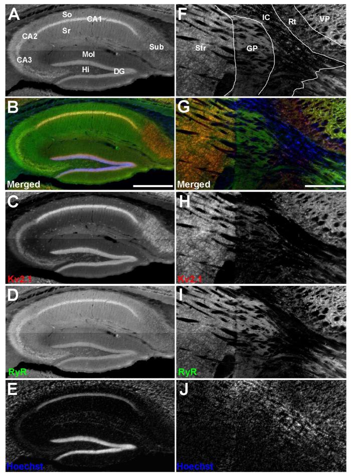
Figure 1.
Distinct patterns of Kv2.1 and RyR coexpression in mouse brain. Mouse brain sections were double immunofluorescence labeled for Kv2.1 (red) and RyR (green), and nuclei were labeled with Hoechst 33258 (blue). Images were acquired with an epifluorescence microscope using an automated stage for mosaic images of brain regions. A-E: Hippocampus. F-J: Striatum and adjacent brain regions. A: Grayscale version of the color image shown in panel B labeled for anatomical regions. B: Merged image of triple immunofluorescence labeling. C: Labeling for Kv2.1. D: Labeling for RyR. E: Labeling for nuclei with Hoechst. F: Grayscale version of the color image shown in panel G labeled for anatomical regions. G: Merged image of triple immunofluorescence labeling. H: Labeling for Kv2.1. I: Labeling for RyR. J: Labeling for nuclei with Hoechst. Labels on panel A: Sub: Subiculum; So: Stratum oriens; Sr: Stratum radiatum; CA1-CA3: respective pyramidal cell layers; Mol: Dentate gyrus molecular layer; HI: Dentate gyrus hilus; DG: Dentate gyrus granule cell layer. Labels on panel F: Str: Striatum; Gp, Globus pallidus; IC, Internal capsule; RT, Reticular nucleus (Thalamic); VP, Ventral posterior nucleus (Thalamic). Scale bar in panel B is 500 μm and is for panels A-E. Scale bar in panel G is 500 μm and is for panels F-J.

