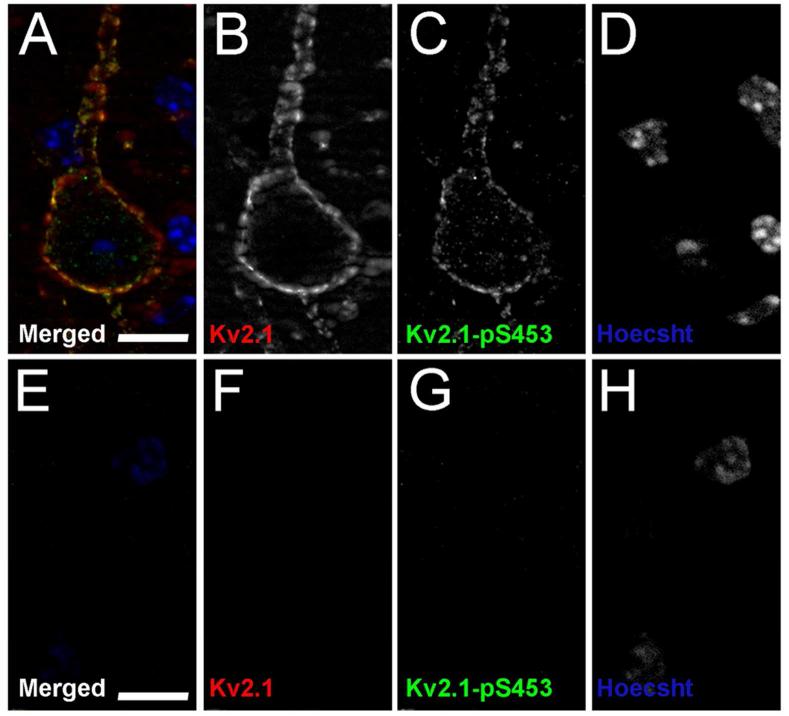
Figure 9. Kv2.1-pS453 validation in Kv2.1-KO mice.
Mouse brain sections from wild type mice, A-D and Kv2.1-KO mice, E-H were double immunofluorescence labeled for general Kv2.1 with the K89/34 mAb (red), and Kv2.1-pS453 (green), and nuclei were labeled using Hoechst (blue). Images of single optical sections of cortical neurons were acquired at equal exposures using an epifluorescence microscope equipped with an ApoTome. Scale bars are 10 μm.

