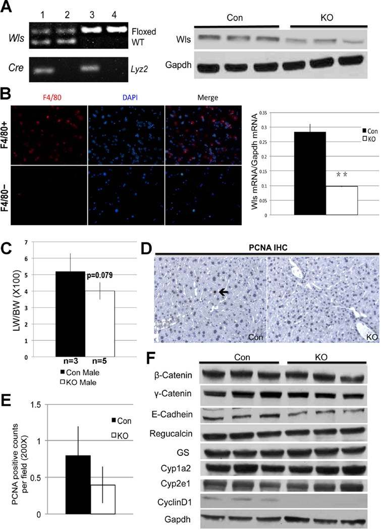
Figure 6. Impact of deletion of Wls in Kupffer cells on hepatic homeostasis.
A. Representative PCR (left) showing genotype of Wls-MKO (Wlsflox/flox; Lyz2-Cre+/−) (lane 3) and Con with genotype Wlsflox/wt; Lyz2-Cre−/− & Wlsflox/flox;Lyz2-Cre−/− (lanes 2 & 4). Representative WB shows a notable decrease in Wls in Wls-MKO liver lysate.
B. Enrichment for Kupffer cells after collagenase perfusion and column separation of non-parenchymal cells is verified by immunocytochemistry on cytospin for F4/80 (red) showing positive staining in the F4/80-positive cell fraction and not in F4/80-negative fraction (left). Wls mRNA expression in F4/80+ cells isolated from Con and Wls-MKO, shows significantly lower expression in the latter (**p<0.001).
C. Bar graph depicts marginally lower LW/BW ratio in Wls-MKO, although difference was not statistically significant.
D. IHC (200×) shows an occasional PCNA-positive hepatocyte in Con at baseline (arrow), while no positive cell is observed in a representative view in Wls-MKO.
E. Quantification of PCNA staining showing less positive cells in Wls-Mac KO than Con at baseline, although differences were insignificant.
F. WB shows no alteration in proteins levels of β-catenin, junctional proteins and pericentral targets. However Cyclin-D1 was notably lower in Wls-MKO livers.

