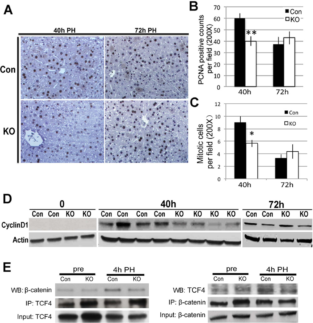
Figure 8. Dampened LR in Wls-MKO after PH.
A. IHC (200×) showing many PCNA-positive hepatocytes in Con livers at 40 hours and 72 hours; however several PCNA-negative hepatocytes were observed in Wls-MKO at 40 hours.
B. Quantification of PCNA staining showing a 33% and significant decease in the number of hepatocytes in S-phase in Wls-MKO at 40 hours (**p<0.01).
C. Quantification of mitotic figures shows a significantly lower numbers in Lrp-LKO as compared at Con at 72 hours after PH. (*p<0.05)
D. Representative WB shows lower Cyclin-D1 levels in Wls-MKO at 40 hours after PH.
E. A representative immunoprecipitation study shows decreased TCF4-β-catenin complex formation at 4 hours after PH in Wls-Mac KO when compared to Con. Immunoprecipitation studies were performed by pull down of either β-catenin or TCF4. Respective input controls are included in analysis as well.

