
Fig. 1.
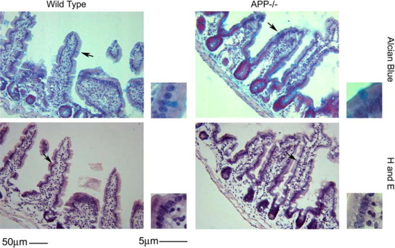
Histology of the small intestine was similar in both APP−/− and wild type controls. Ileum tissue samples were collected from C57BL6/J wild type and APP−/− mice, fixed in 4% paraformaldehyde, serially sectioned, and stained using routine histology H & E (hematoxylin and eosin) and Alcian blue. Arrows indicate the location of the region of interest shown as an enlarged inset to the right of each panel. Representative images from 5 animals per condition are shown.