
Fig. 4.
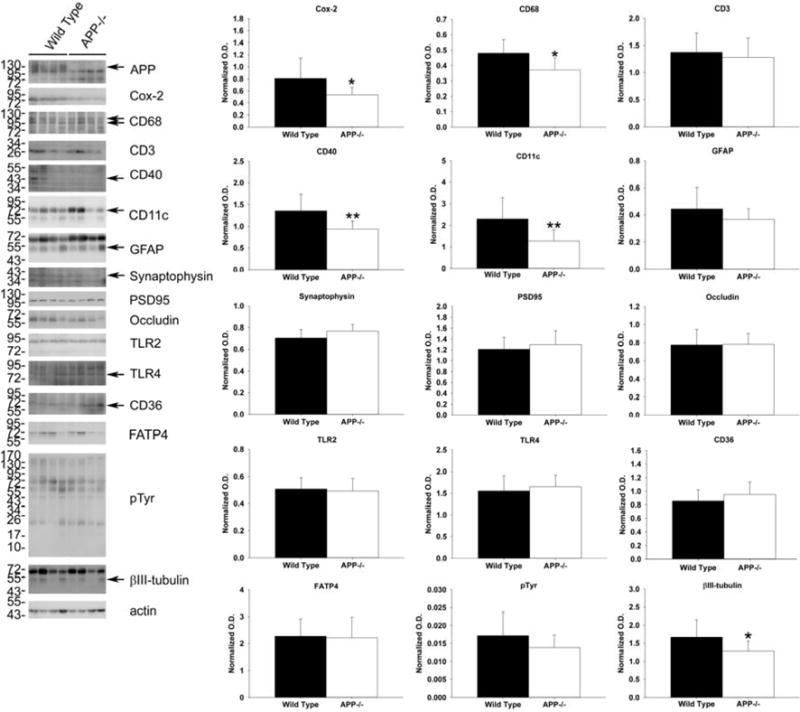
Western blot analysis of cox-2, CD68, CD40, CD11c and βIII-tubulin demonstrated decreased protein levels in the small intestine of APP−/− compared to wild type mice. Ileum of the small intestine was collected from APP−/− and C57BL6/J wild type mice.. The tissue was lysed, resolved by 10–15% SDS-PAGE and Western blotted using anti-CD68, CD3, CD40, CD11c, GFAP, TLR2, TLR4, CD36, FATP4, pTyr synaptophysin, PSD95, APP, cox-2, occludin, βIII tubulin (neuronal loading control), or actin (general loading control) antibodies. Antibody binding was visualized by chemiluminescence. Blots from 4 of the animals for each strain are shown. Optical densities of the Western blotted ileum proteins were normalized against their respective actin (cox-2, CD68, CD3, CD40, CD11c, GFAP, TLR2, TLR4, CD36, FATP4, pTyr and occludin) or βIII tubulin (synaptophysin and PSD95) loading controls and averaged (+/−SD) from 10 animals per each condition *p<0.05.