
Figure 3.
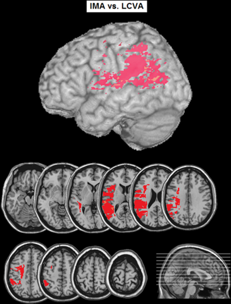
Voxel-based lesion analysis comparing left-hemisphere stroke patients with ideomotor apraxia (IMA) to left hemisphere stroke patients without IMA (control). Colored voxels indicate areas where the percentage of participants with damage to a given voxel was greater in the IMA than control group.