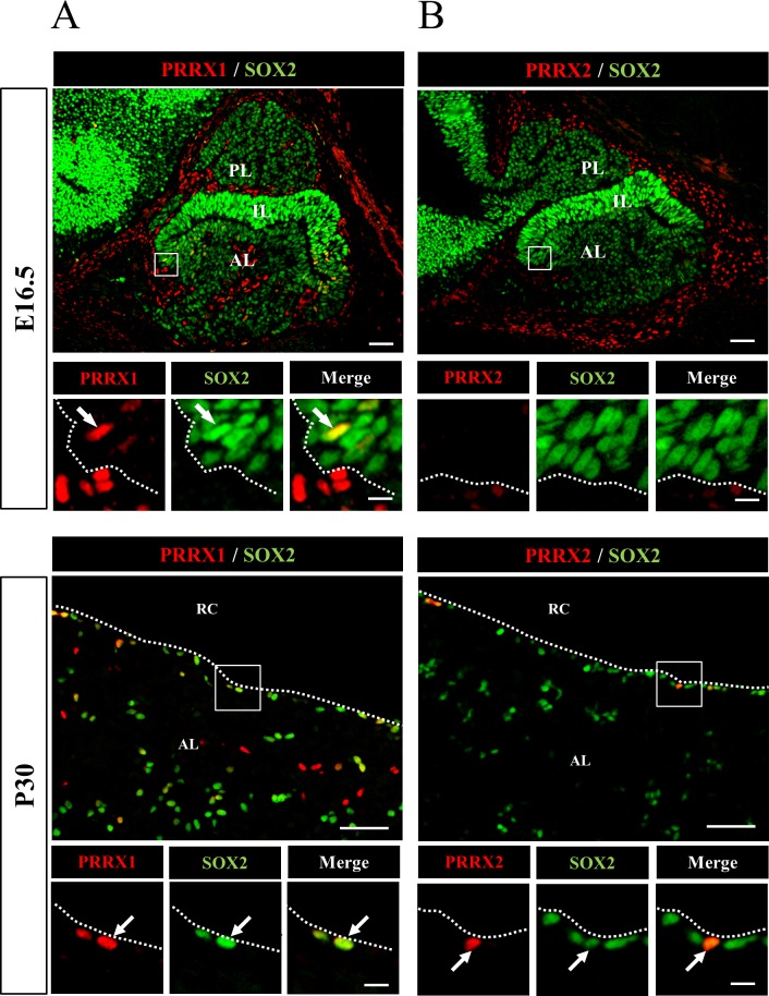
Fig. 1.
Localization of PRRX1 and PRRX2 in pituitary stem/progenitor cells. Immunohistochemistry for PRRXs (A, PRRX1; B, PRRX2) and SOX2 was performed using frozen sections of rat pituitary at embryonic day 16.5 (E16.5) and postnatal day 30 (P30). Areas of PRRXs and SOX2 in open boxes, which were visualized with Cy3 (red) and fluorescein isothiocyanate (green), were enlarged as shown below together with the merged image. The arrow and dotted line indicate cells double positive for PRRXs and SOX2 and the marginal cell layer, respectively. AL, anterior lobe; IL, intermediate lobe; PL, posterior lobe; RC, Rathke’s cleft. Scale bars: 50 µm and 10 µm (enlarged images).

