Fig. 3.
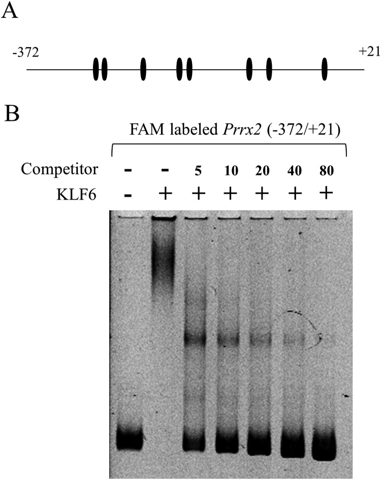
Electrophoretic mobility shift assay (EMSA) for KLF6. (A) A diagram of the 5’ upstream (–372/+21) region of Prrx2, which was used as a binding probe, is shown. A putative binding site (CCNCNCCN including GC element and CACCC) of KLF6 is shown with a closed ellipse. (B) EMSA was performed using a 100 fmol FAM-labelled fragment (–372/+21) without and with a 5–80 molar excess amount of non-labelled fragment (–372/+21) as a competitor to confirm the specific DNA/protein complex. Electrophoresis of the FAM-labelled fragment (probe) alone is shown at the left of the panel.

