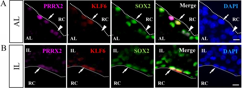
Fig. 5.
Immunohistochemistry of KLF6 together with PRRX2 and SOX2. Immunohistochemistry for KLF6 was performed using frozen sections of rat pituitary at P20. Note that immunohistochemical images of KLF6, PRRX2 and SOX2 were visualized with Cy3 (red), Cy5 (purple) and fluorescein isothiocyanate (green) and counterstained with DAPI (blue). Dotted lines indicate the marginal cell layer (MCL). AL, anterior lobe; IL, intermediate lobe; RC, Rathke’s cleft. Scale bars: 10 μm.

