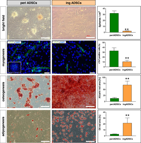
Figure 3.

Comparison of differentiation potential of two sources of ADSCs. After cardiac induction, both ADSCs showed enlarged morphology, but the pericardial ADSCs (periADSCs) formed spherical structures similar to cardiospheres and significantly more cTnT-expression cells in comparison with inguinal ADSCs (ingADSCs, n = 8 in each group). The inset shows the formation of cTnT-expressing cells within the spherical structure. In contrast, periADSCs were less competent to adipogenic (P < 0.01; n = 5 in each group) and osteogenic differentiation (P < 0.01; n = 5 in each group), as indicated by oil red and alizarin staining. Bar = 100 μm.
