Figure 1.

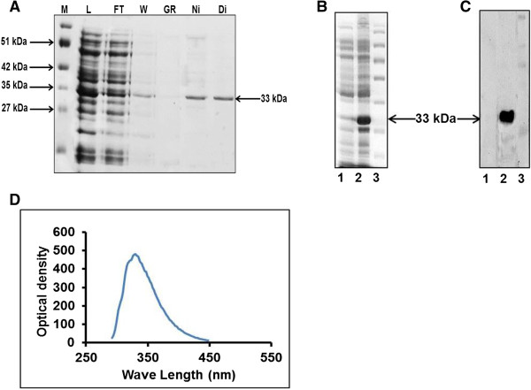
SDS-PAGE profile of buffalo HOXC11 protein. A) SDS-PAGE (15%) for HOXC11 protein showing its resolved chromatographic fractions. M denotes marker, L: load, FT: flow through, W: wash, Gr: gradient, Ni: Ni-NTA fraction and Di: dialyzed protein. B) SDS-PAGE of HOXC11 protein; lane 1 represents uninduced; lane 2, induced and lane 3, marker. C) Validation of purified buffalo HOXC11 protein by Western blot with anti-His and HRP conjugated anti-rabbit IgGs used as primary and secondary antibodies, respectively. Lane 1, represents uninduced; lane 2, induced and lane 3, marker. D) Tryptophan emission spectra. Fluorescence of refolded protein was scanned at 280 nm excitation wavelength and 300–400 nm emission wavelength.
