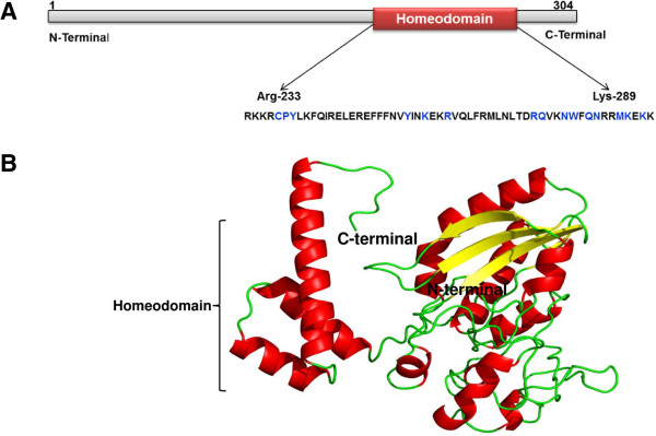
Figure 8.

Modeled 3D structure of the buffalo HOXC11 protein. A) Diagrammatical representation of homeodomain (56 aa long) and possible binding site residues (in blue) of HOXC11 protein. B) The 3D structure of buffalo HOXC11 was generated by I-TASSER and visualized by PyMol (version 1.7). Helix, beta sheets and coils/loops are shown in red, yellow and green, respectively.
