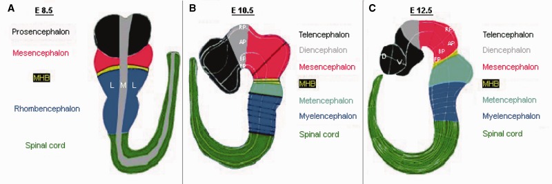
Figure 1.
CNS regionalization of the mouse embryo at different developmental stages as used for the database structure. (A) Developmental stage E8.5: The whole embryo is divided into five regions along the anterior–posterior axis: prosencephalon, mesencephalon, MHB, rhombencephalon, spinal cord; along the medial–lateral axis the embryo is divided into medial and lateral, and the region in between is considered as mediolateral boundary. (B) Developmental stage E10.5: The embryo regionalization along the anterior–posterior and along the dorsal–ventral (previous medial–lateral) axis is as follows: telencephalon, diencephalon, mesencephalon, MHB, metencephalon (r1), myelencephalon (r2-r8) and spinal cord; for all CNS regions, the new regionalization along the dorsal–ventral axis is RP, AP, ABB, BP and FP. (C) Developmental stage E12.5: The mouse embryo is regionalized along the anterior–posterior axis as follows: telencephalon (anterior, posterior), diencephalon (anterior hypothalamus, posterior hypothalamus, prethalamus, thalamus), mesencephalon (anterior, posterior), MHB (anterior, posterior), metencephalon (r1), myelencephalon (r2-r8) and spinal cord (cervical, thoracic, lumbar, sacral, caudal); the telencephalon, diencephalon and MHB are subdivided along the dorsal–ventral axis into RP, AP, ABB, BP and FP; the mesencephalon, metencephalon and myelencephalon are subdivided into dorsal and ventral regions and/or neuronal populations along the dorsal–ventral axis; the spinal cord is subdivided into roof plate, dl1, dl2, dl3, dl4, dl5, dl6, v0, v1, v2, v3, v4, mn and floor plate, where dl1 to dl6 describe the dorsal interneurons, and v0 to v3 and mn denote the ventral interneurons (not shown).

