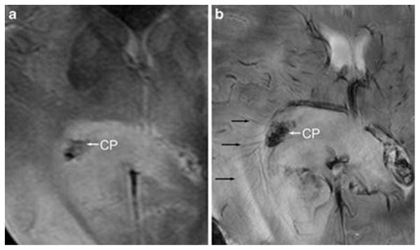
Fig. 1.

MR images of an axial slice of a human brain at (a) 1.5 and (b) 7 T. The image acquired at 7 T enables visualization of blood vessels (black arrows) and choroid plexus (CP, an abnormality in the right lobe) that are not clearly visible at 1.5 T.7 Reprinted from C. Moenninghoff, S. Maderwald, J. M. Theysohn, O. Kraff, M. E. Ladd, N. El Hindy, J. van de Nes, M. Forsting and I. Wanke, Imaging of Adult Astrocytic Brain Tumours with 7 T MRI: Preliminary Results, Eur. Radiol., 2010, 20, 704–713, with kind permission from Springer Science and Business Media.
