Figure 2:


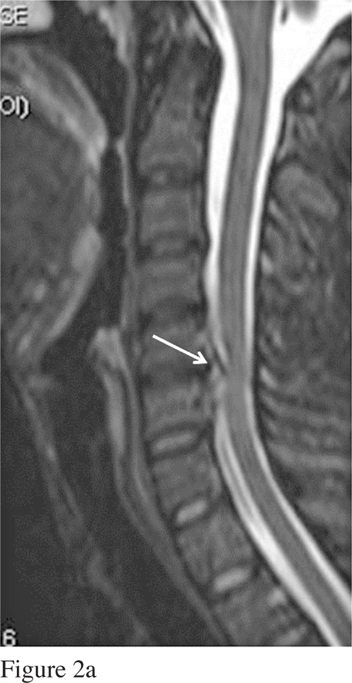
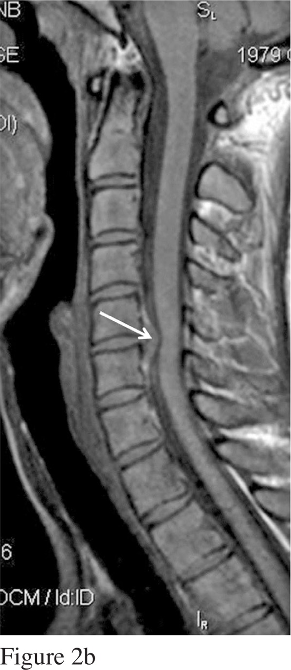
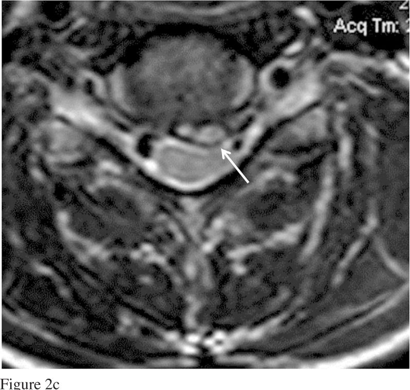
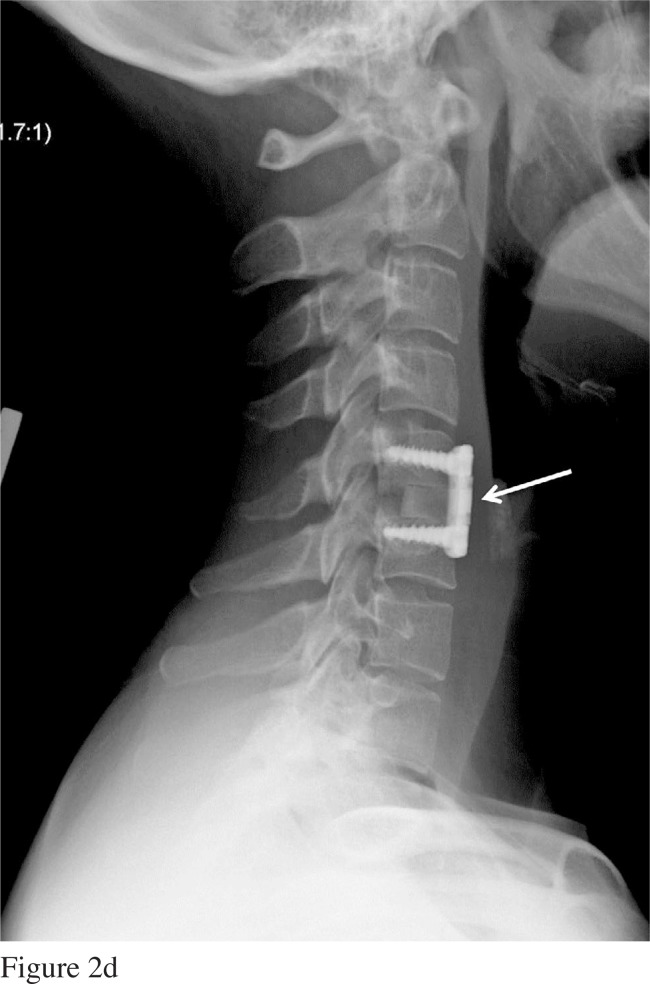
(a) T2-weighted sagittal MRI scan of patient’s cervical spine (2011) demonstrating significant right paracentral herniated nucleus pulposus of the C5–6 intervertebral disc (white arrows in all figures). (b) Sagittal T1 MRI of cervical spine depicting large C5–6 disc herniation and elevation of posterior longitudinal ligament, (c) Axial image of the same C5–6 disc as in (b), (d) Plain film radiographs of patient’s cervical spine post C5–6 anterior cervical decompression and fusion. Note interbody bone graft and plate affixed to the anterior aspect of the cervical spine (white arrow).


