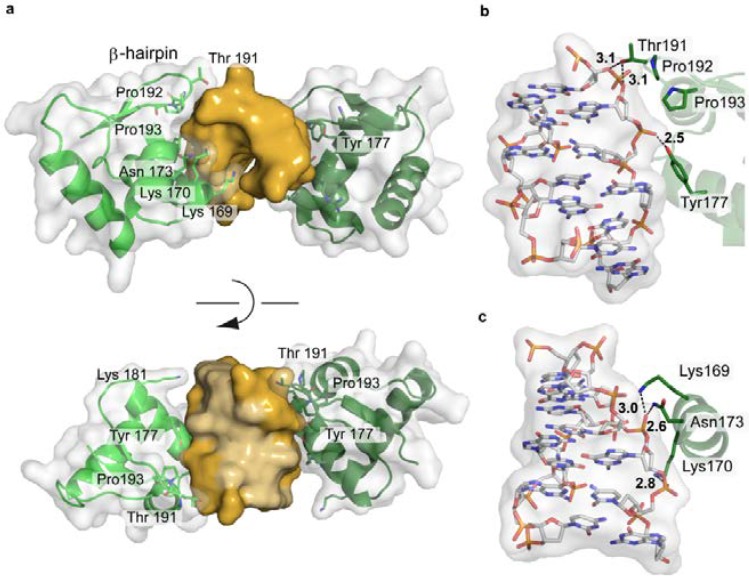
Figure 13.
Structure of the Zα-domain of ADAR1 bound to left-handed Z-DNA (PDB code 3IRQ). (a) Schematic representation of the protein-DNA complex, highlighting the surface fit of the two macromolecules. The DNA is shown in gold, with the two bound proteins in green, overlaid with their semi-transparent surface; (b,c) Electrostatic interactions between ADAR1 and the Z-DNA backbone.

